Blood Vessels Labeled Simple - Heart Blood Flow Circulation Anatomical Diagram With ... - Blood vessels are an integral component of the circulatory system.
byAdmin-
0
Blood Vessels Labeled Simple - Heart Blood Flow Circulation Anatomical Diagram With ... - Blood vessels are an integral component of the circulatory system.. Does not cover the pathology content. August 17, 2020 so, you want to learn. What do you mean by the term dlapedesls? Blood flows throughout the body tissues in blood vessels, via bulk flow (i.e., all constituents together and in one direction). This is a free printable worksheet in pdf format and holds a printable version of the quiz blood vessel labeling.
Simple squamous epitheliumendothelium in blood vessels blood vessel color images Dimitrios mytilinaios md, phd • last reviewed: Download pdf worksheet blank download pdf worksheet labeled consolidate your knowledge with interactive quizzes. • identification of blood vessels as arteries, capillaries or veins from the structure of their walls. While most blood vessels are located deep from the surface and.
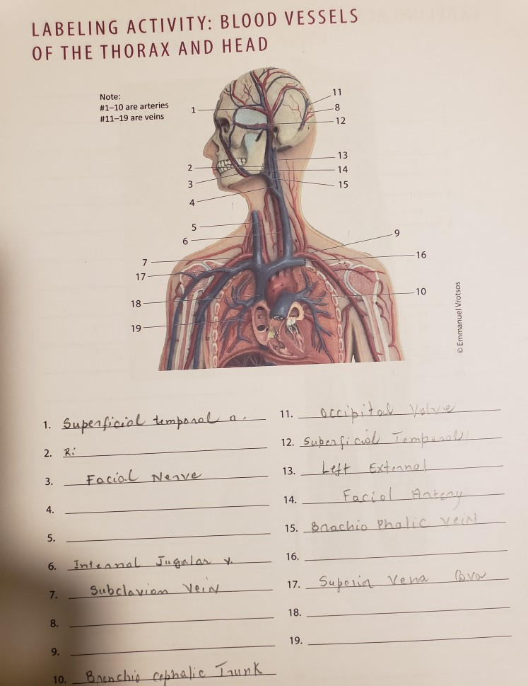
Solved: LABELING ACTIVITY: BLOOD VESSELS OF THE THORAX AND ... from media.cheggcdn.com The inner lining is the endothelium and is surrounded by subendothelial connective tissue. The most important types, arteries and veins, carry all blood vessels have the same basic structure. Allows diffusion of gases and nutrients from blood into the body cells. Start studying blood vessels labeling. Now that we understand the structure and function of the heart, which is to pump blood all around the body, what is the blood traveling around in exactly? Hma practical 3 virtual slides. By printing out this quiz and taking it with pen and paper creates for a good. Which of the labeled layers in the diagram of the arterial wall is composed of a simple squamous epithelium, a basement membrane and a layer of.
Blood vessels (labeled) coloring page.
Blood vessels are referred to collectively as the vascular system and, together with the heart, make up the circulatory system or cardiovascular system. Does not cover the pathology content. The iliac, femoral, popliteal and tibial (calf) veins are the deep veins in the legs. These vessels transport blood cells, nutrients, and oxygen to the tissues of the body. The inner lining is the endothelium and is surrounded by subendothelial connective tissue. It is made up of. August 17, 2020 so, you want to learn. Hma practical 3 virtual slides. Blood vessels are an integral component of the circulatory system. Ready to learn about the blood vessels of the abdomen and pelvis (the abdominopelvic blood vessels)? They also take waste and carbon dioxide away from the tissues. When chemoreceptors in blood vessels detect high levels of carbon dioxide in the blood, they stimulate all of the following changes except. This page is about blood vessel histology slide labeled,contains artery microscope slide.
Hma practical 3 for monday july 23 and wednesday july 25. They also take waste and carbon dioxide away from the tissues. Blood vessels are intricate networks of hollow tubes that transport blood throughout the entire body. Each pixel must be labeled 1 if it is part of a blood vessel in the image, and 0 if not. Allows diffusion of gases and nutrients from blood into the body cells.
Artery and Vein DIAGRAM | Blood vessels anatomy, Arteries ... from i.pinimg.com These vessels transport blood cells, nutrients, and oxygen to the tissues of the body. Deep veins, located in the center of the leg near the leg bones, are enclosed by muscle. This page provides histology support information for blood vessel structure. All blood vessels are specifically structured to perform their function. Hma practical 3 for monday july 23 and wednesday july 25. After passing through the aortic hiatus (t12), it descends slightly to the left of the lumbar vertebrae, with the inferior vena cava being just. Retinal_blood_vessel_segmentation.ipynb this is a simple implementation neural net with. This page is about blood vessel histology slide labeled,contains artery microscope slide.
Deep veins, located in the center of the leg near the leg bones, are enclosed by muscle.
Blood vessels are intricate networks of hollow tubes that transport blood throughout the entire body. • identification of blood vessels as arteries, capillaries or veins from the structure of their walls. Molly smith dipcnm, mbant • reviewer: Blood flows throughout the body tissues in blood vessels, via bulk flow (i.e., all constituents together and in one direction). Simple squamous epitheliumendothelium in blood vessels blood vessel color images Blood vessels are flexible tubes that carry blood, associated oxygen, nutrients, water, and hormones throughout the body. Blood vessels are referred to collectively as the vascular system and, together with the heart, make up the circulatory system or cardiovascular system. Hma practical 3 virtual slides. Dimitrios mytilinaios md, phd • last reviewed: The blood vessels are the components of the circulatory system that transport blood throughout the human body. Differentiate among the structure of arteries, veins, and capillaries. Transcribed image text from this question. Download pdf worksheet blank download pdf worksheet labeled consolidate your knowledge with interactive quizzes.
Blood vessels cannot function properly when inhibited by vascular diseases. These vessels transport blood cells, nutrients, and oxygen to the tissues of the body. The intima is a simple epithelium made up of a single layer of flat epithelial cells. The iliac, femoral, popliteal and tibial (calf) veins are the deep veins in the legs. The blood vessels are part of the circulatory system and function to transport blood throughout the body.
Blood Vessels: Arteries, Capillaries & More - Video ... from study.com Blood flows throughout the body tissues in blood vessels, via bulk flow (i.e., all constituents together and in one direction). Hma practical 3 for monday july 23 and wednesday july 25. Blood vessels are vital for the body and play a key role in diabetes helping to transport glucose and insulin. Start studying blood vessels labeling. The inner lining is the endothelium and is surrounded by subendothelial connective tissue. ⇒ click on the diagram to show / hide labels. Structure of blood vessels in general, blood vessels have a walls composed of three layers as follows: The vessels narrow, reducing blood flow, and become less responsive to stimuli inducing vascular regression.
They include arteries, veins, and capillaries.
In the label the blood vessels activity, students analyze the drawing of the human body and label the arteries that are indicated in the picture. Hma practical 3 for monday july 23 and wednesday july 25. What do you mean by the term dlapedesls? By printing out this quiz and taking it with pen and paper creates for a good. The iliac, femoral, popliteal and tibial (calf) veins are the deep veins in the legs. Carry blood towards the heart (usually deoxygenated blood, except for the pulmonary vein). Allows diffusion of gases and nutrients from blood into the body cells. Blood flows throughout the body tissues in blood vessels, via bulk flow (i.e., all constituents together and in one direction). Molly smith dipcnm, mbant • reviewer: An extraordinary degree of branching of blood vessels exists within the human body, which ensures that nearly every cell in the body lies within a short distance from at least one of. They include arteries, veins, and capillaries. Related posts of the human blood vessels labeled digestive system free online quiz blood vessel labeling there are five main types of blood vessels: Blood vessels are intricate networks of hollow tubes that transport blood throughout the entire body.
After passing through the aortic hiatus (t12), it descends slightly to the left of the lumbar vertebrae, with the inferior vena cava being just blood vessels labeled. Each pixel must be labeled 1 if it is part of a blood vessel in the image, and 0 if not.